 |
|
ENFERMEDAD DE QUERVAIN |
|
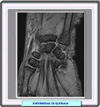
LThe coronal T2-weighted MRI image sequence showed abnormal segmental tendinous thickening involving the first dorsal extensor component of the wrist just proximal the radial styloid process, with a mild amount of fluid within the tendon sheath. There was no evidence of significant bony abnormality or soft-tissue masses or collections. These findings are consistent with a diagnosis of de Quervain tenosynovitis, an inflammatory process that typically involves the first dorsal extensor components of the wrist (namely, the abductor pollicis longus [APL] and the extensor pollicis brevis [EPB]) within the narrow fibro-osseous tunnel through which they normally pass. Although MRI is not typically necessary for the diagnosis of de Quervain tenosynovitis, the image here nicely shows the inflammatory changes characteristic of the condition. De Quervain tenosynovitis was first described in 1895 by a swiss surgeon, Fritz de Quervain, who reported 5 cases of patients with a tender, thickened first dorsal wrist compartment. The condition has subsequently named after him. It is considered to be the second most common entrapment tendinitis of the hand and wrist (after trigger digit syndrome). In 1930, Finkelstein reviewed the literature and reported 24 additional cases. He felt that chronic trauma should be considered as the principal cause of de Quervain tenosynovitis. From his work, the author derived the well-known "Finkelstein sign", which is used to diagnose the disease. The Finkelstein sign classically occurs in mothers of infants 6-12 months of age. Interestingly, the cause is believed to be principally endocrine and related to fluid retention in breast-feeding mothers, not solely as a result of repetitive lifting motion. De Quervain tenosynovitis has also, however, been described in fathers. This suggests that the condition can occur in the absence of postpartum endocrine changes. Repetitive trauma in manual laborers is also a common cause. The pain itself may appear either gradually or suddenly.[1,2,11] In most cases, the patient will often complain of weeks to months of gradually worsening pain proximal and posterior to the radial styloid process, especially when using the thumb. The patient will experience difficulties with gripping and pinching and, in severe cases, the affected hand may be too painful to use. The pain may radiate into the thumb, or it may radiate proximally into the ipsilateral forearm or shoulder. In the vast majority of the cases, a discrete soft-tissue mass corresponding to the affected tendon sheath thickening of the first extensor compartment at the level of the distal dorsolateral aspect of the radial styloid process is encountered on clinical examination. The presence of multiple tendon slips and variable insertions of the abductor pollicis longus, and the presence of a separate compartment for the extensor pollicis brevis, have been noted in patients who have de Quervain tenosynovitis. Anatomical studies involving human cadavers have shown great variability in the incidence of separate compartments for the extensor pollicis brevis and abductor pollicis longus tendons. In a large series, there was a separate compartment for the extensor pollicis brevis in 120 (40%) of 300 dissected wrists. In addition, several reports have described the anatomical variations of the first dorsal wrist compartment, and suggested that normal variations of the tendons of this compartment could be related to a cause of de Quervain tenosynovitis. No associated skin changes or discoloration are typically present.[3,4,5,11] De Quervain tenosynovitis is diagnosed clinically and rarely requires laboratory testing or imaging for confirmation (such as occurred in this case); however, imaging abnormalities have been described, and MRI is currently the preferred imaging modality. It is most often ordered in atypical cases in which an alternative diagnosis is being entertained, or in cases that are recurrent or resistant to conventional treatment. An MRI typically shows thickening and edema of the involved segment of the tendon, which is best depicted on the long TR sequences (mainly T2-weighted images). There may be hyperintense fluid signal-intensity surrounding or within the affected tendon sheath. A normal caliber tendon does not exclude the diagnosis. Intravenous (IV) administration of gadolinium is not usually necessary; however, when administered, enhancement within the tendons and surrounding soft tissue consistent with tenosynovitis may be seen. Additionally, in atypical cases, an MRI scan is helpful in excluding other etiologies of wrist pain, such as ganglion cysts and nerve sheath tumors.[6,11] Several studies have shown that ultrasonography is also a reliable and noninvasive method for detecting tenosynovitis changes. Ultrasonography of the symptomatic tendon typically shows distention in the tendon sheath, with surrounding fluid that is hypoechoic or anechoic. An axial scan of the tendon will exhibit a so-called "double target" appearance. Ultrasonography is also used to guide the delivery of therapeutic agents into the affected tendon sheath (as injections into the tendon should be avoided) and to follow the response to treatment. It can also help to determine the segment with the most inflammatory changes, allowing for precise administration of the steroids. Studies have shown a significant decrease in the thickness of the affected tendon sheath at 1 week after a local corticosteroid injection, with complete relief of the clinical symptoms and signs observed at 6 and 12 weeks postinjection. Conventional radiography is typically of limited usefulness, but it may show the area of soft-tissue swelling proximal to the styloid process. Some clinical series have shown focal radial styloid process abnormality at the first dorsal wrist compartment that is significantly associated with de Quervain tenosynovitis. Moreover, in clinical conditions that may mimic de Quervain tenosynovitis, radiographs can be helpful in ruling out offending bony pathology, such as degenerative bony changes.[9,10] Histologic examinations may reveal myxoid degeneration, which is responsible for the thickening observed in the sheath, and intramural deposits of mucopolysaccharides predominantly within the subsynovial regions. Macroscopically, classic de Quervain tenosynovitis demonstrates chronic inflammation, with scar formation and stenosis of the approximately 1-cm section of the fibro-osseous tunnel of the first dorsal compartment.[7] The goal of treatment for de Quervain tenosynovitis is to relieve the pain caused by irritation and swelling. Treatment includes conservative measures such as splinting of the affected thumb and wrist, which is sometimes ineffective (pain may return as soon as the splint is discontinued). In most cases, however, the disease is largely self-limiting and usually resolves after cessation of breast feeding. Injection of corticosteroids into the tendon sheath usually results in pain relief in about 82-95% of the cases. Symptomatic improvement from corticosteroid injection is usually observed within a few days after injection. The prognosis is generally good. A patient will generally return to full function after the inflammation quiets down with treatment. If 4-6 months of conservative treatment fails to alleviate the symptoms, then surgical release of the first dorsal compartment may be considered. The procedure is usually done on an out-patient basis. The surgery typically involves identification and cutting of the diseased tendon sheath segment under local anesthesia. Patients commonly return to their normal activities within 2-3 weeks. The procedure has been reported to be successful in about 90% of the cases.[4,8] The patient in this case failed conservative treatment (approximately 4 weeks) with splinting; there was only very mild improvement of her symptoms. As a result, intralesional injection of steroid using a 25 gauge needle was performed. The patient regained function and experienced pain relief within 3-4 days following the injection. No symptom recurrence was reported.
|
REFERENCIAS
|
|


